


Page 15 - 南京医科大学自然版
P. 15
第46卷第10期             王新新,黄伯贤,陆嘉逢. PUS7通过mRNA假尿嘧啶化调控胚胎发育过程中神经
                 2026年10月             谱系分化平衡[J]. 南京医科大学学报(自然科学版),2026,46(10):1437-1449                 ·1445 ·


                           [9]
                或表达维持 ,这类同步变化的基因更可能反映                             联。因此,本研究重点关注Ψ修饰下降且mRNA表达
                Pus7缺失后Ψ修饰降低与转录本表达受损之间的关                          下调的交集基因,以筛选可能受PUS7介导mRNA Ψ
                  A   d 0              d 2   d 3         d 5                B         WT           Pus7⁃KO

                            N2B27            N2B27+RA
                                                                                d 1
                                         Neuronal differentiation
                             Stage 1          Stage 2
                  C    Pluripotency     Neuroectoderm⁃related genes
                                                                                d 3
                     d 0
                                                                   log2 FC
                                                                  (KO/WT)
                     d 3                                             2
                                                                     0
                        Klf4  Sox2  Pou5f1  Nanog  Ncam1  Sox9  Map2k6  Map2  En2  Nptx2  Hes5  Aldh1l1  Otx1  Serf1  Nr2f1  Meis1  Sox3  Edn3  Irx2  Mkx6⁃1  Meis2  Gli3  Prdm1  Meis3  Pax2  Phox2b  Nsg2  Zic5  En1  Pax6  Six3  Sox1  Pax3  Olig2  -2  d 5


                D      WT                  WT                 WT                 WT                 WT
                       Pus7⁃KO             Pus7⁃KO            Pus7⁃KO            Pus7⁃KO            Pus7⁃KO
                   40                  25                 15                  8          ***    1.5
                                               ***
                  mRNA level  30      Sox1 mRNA level  15  mRNA level  10  **  ***  mRNA level  6 4  Aldh1l1 mRNA level  1.0
                                       20
                           ***
                   20
                  Pax6  10             10 5  **          Map2  5             Ncam1  2  *        0.5

                    0                   0                  0                  0                  0
                        d0  d3  d5         d0  d3  d5          d0  d3  d5         d0  d3  d5         d0  d3  d5
                E                        F                                              G
                        WT  Pus7⁃KO                  Pus7⁃KO                WT             3.0×10 3
                                                                                                          WT
                      d0 d3 d5 d0 d3 d5         Merge      SOX1      Merge      SOX1
                                                                                                          Pus7⁃KO
                 PAX6               50 kDa                                                 2.0×10 3
                                                                                           Count
                GAPDH               36 kDa  0
                                            d                                                   3
                                                                                           1.0×10
                         PAX6  *   WT                                                           0  3    3   4  5
                  protein level 40  Pus7⁃KO  3  d                                       H       -10 0 10  10  10
                          **
                                                                                                     SOX1⁃FITC
                   30
                   20
                                                                                                     ***
                  PAX6  10                                                                     (of DAPI + )  50
                                                                                               40
                    0
                      d0  d3  d5            5                                                  cells 30
                                                                                               20
                                            d
                                                                                               % FITC +  10 0
                                                                                                   WT Pus7⁃KO
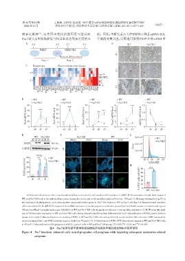
                   A:Schematic illustration of the experimental workflow for directed neural ectoderm differentiation of mESC. B:Representative bright⁃field images of
                WT and Pus7⁃KO cells at the indicated time points during directed neural ectoderm differentiation(Scale bar=100 μm). C:Heatmap showing the log 2 FC in
                the expression of pluripotency⁃ and neuroectoderm⁃associated marker genes in Pus7⁃KO relative to WT at day 0 and day 3 of directed neural ectoderm
                differentiation(n=3). D:qRT⁃PCR analysis of the mRNA expression of neural progenitor cellmarker genes(Sox1 and Pax6),mature neuronal marker genes
               (Ncam1 and Map2)and glial marker gene(Aldh1l1)in WT and Pus7⁃KO cells during directed neural ectoderm differentiation(n=3). E:Western blot anal⁃
                ysis of PAX6 protein expression in WT and Pus7⁃KO cells during directed neural ectoderm differentiation(n=3). Quantification of PAX6 protein levels is
                shown on the right. F:Immunofluorescence staining of SOX1 in WT and Pus7⁃KO cells during directed neural ectoderm differentiation;DAPI was used for
                nuclear staining(blue),and SOX1 is shown in green. Scale bar=50 μm(n=3). G:Distribution of SOX1⁃FITC fluorescence signals in WT and Pus7⁃KO cells
                                                                                             ***
                                                                                   **
                                                                             *
                at d3(n=3). H:Quantification of the proportion of SOX1⁃positive cells in WT and Pus7⁃KO groups. P < 0.05,P < 0.01,and P < 0.001.
                                  图4   Pus7敲除促进早期神经祖细胞程序增强并伴随后续成熟相关程序受阻
                Figure 4  Pus7 knockout enhanced early neural progenitor cell programs while impairing subsequent maturation⁃related
                        programs

