


Page 53 - 南京医科大学自然版
P. 53
第46卷第10期             刘怡珂,李超凡,宋海燕,等. 鞣花酸靶向miR⁃18a/TSP⁃1轴减轻缺血性脑损伤[J].
                 2026年10月                    南京医科大学学报(自然科学版),2026,46(10):1479-1491                     ·1483 ·


                2.3  EA在MCAO模型中发挥神经保护作用并改善                        处理加速了缺血性脑损伤后的功能恢复(图 3A)。
                神经功能                                              组织病理学支持上述行为学结果,HE 染色显示,
                    从第 3 天开始,EA 预处理组 Longa 评分显著低                  MCAO组大鼠缺血半暗带脑组织出现明显的神经元
                于MCAO组(P < 0.05),且趋势持续存在,表明EA预                    排列紊乱、胞体固缩及空泡化,而EA预处理能显著
                A                            B          Bright⁃field          TUNEL                Merge
                   1.5
                                  **
                               *
                  Cell activity  1.0      *     Control


                   0.5


                    0                           OGD
                     10 μmol/L EA 50 μmol/L EA 100 μmol/L EA
                     Control
                               75 μmol/L EA
                        25 μmol/L EA
                C
                     100
                              **
                      80                        μmol/L EA
                     (%)  60      ** △          25
                     TUNEL  40       * △△


                      20
                       0                        μmol/L EA
                        Control 25 μmol/L EA    50
                                50 μmol/L EA
                             OGD

                D           Control                  OGD                  25 μmol/L EA           50 μmol/L EA
                     10 7  Q1⁃1    Q1⁃2      10 6  Q1⁃1     Q1⁃2     10 6  Q1⁃1    Q1⁃2      10 6  Q1⁃1    Q1⁃2
                                              9.72%
                                   0.17%
                      6.47%
                                                            7.16%
                                                                      9.31%
                                                                                   5.56%
                     10 6                    10 5                    10 5                    10 5  8.05%   4.70%
                  PerCP⁃H  10 5  10 4      PerCP⁃H  10 4           PerCP⁃H  10 4          PerCP⁃H  10 4
                     10 3  Q1⁃3    Q1⁃4      10 3  Q1⁃3     Q1⁃4     10 3  Q1⁃3    Q1⁃4      10 3  Q1⁃3    Q1⁃4
                     10 2  92.93%  0.43%     10 1.9  74.93%  8.19%   10 1.9  81.49%  3.63%   10 1.9  85.84%  1.40%
                     10 2.7  10 4  10 5  10 6  10 2.9  10 5  10 7  10 9  10 3.1  10 5  10 7  10 9  10 3.1  10 5  10 7  10 9.3
                           FITC⁃H                   FITC⁃H                  FITC⁃H                  FITC⁃H
                E                     F                           G                      H
                  20                                                   1.0                   0.8     **
                                                           50 μmol/L EA
                 (%)  15  **                 Control  OGD  25 μmol/L EA  protein expression  0.8  ** △  * △△  0.6  ** △  * △△
                 Apoptosis rate  10 5  ** △  * △△  Bcl⁃2      26 kDa   0.4     **           Bax protein expression  0.4
                                                                       0.6

                                                              21 kDa
                                        Bax
                                                                                             0.2
                                      β⁃actin                 42 kDa  Bcl⁃2  0.2
                   0                                                    0                     0
                   Control 25 μmol/L EA                                 Control 25 μmol/L EA  Control 25 μmol/L EA
                                                                             OGD
                        OGD
                                                                                                    OGD
                                                                                                       50 μmol/L EA
                                                                                50 μmol/L EA
                           50 μmol/L EA
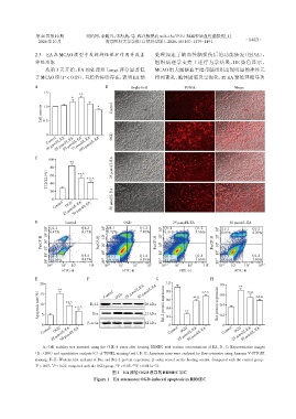
                   A:Cell viability was assessed using the CCK⁃8 assay after treating RBMEC with various concentrations of EA. B,C:Representative images
               (B,×200)and quantitative analysis(C)of TUNEL staining(red). D,E:Apoptosis rates were analyzed by flow cytometry using Annexin V⁃FITC/PI
                staining. F-H:Western blot analysis of Bax and Bcl⁃2 protein expression. β⁃actin served as the loading control. Compared with the control group,
                *      **                         △      △△
                P < 0.05,P < 0.01;compared with the OGD group,P < 0.05, P < 0.01(n=3).
                                                图1   EA减轻OGD诱导的RBMEC凋亡
                                        Figure 1 EA attenuates OGD⁃induced apoptosis in RBMEC

