


Page 54 - 南京医科大学自然版
P. 54
第46卷第10期
               ·1484 ·                           南 京    医 科 大 学 学         报                        2026年10月


              A                                                                  B     100
                   Bright⁃field      Hoechst          EdU            Merge
                                                                                       80
               Control                                                                (%)  60      ** △  * △△

                                                                                      EdU  40  **

                                                                                       20
               OGD                                                                      0
                                                                                         Control 25 μmol/L EA
                                                                                              OGD
                                                                                                 50 μmol/L EA
               μmol/L EA
                                                                                                       50 μmol/L EA
               25                                                               C        Control  OGD  25 μmol/L EA
               μmol/L EA                                                        Cyclin D1                34 kDa
                                                                                  PCNA
                                                                                                         29 kDa
               50                                                                 β⁃actin                42 kDa
              D                                 E                                  G
                                                     0.8         *   △△                  400
                    Cyclin D1 protein expression  0.3  **  *  * △  PCNA protein expression  0.4  **  Cell migration number  300  **  ** △  △△
                     0.4
                                                     0.6
                                                                                         200
                     0.2
                                                     0.2
                     0.1
                                                                                         100
                      0
                                                                                                OGD
                                                              50 μmol/L EA
                                                           OGD
                      Control 25 μmol/L EA            0 Control 25 μmol/L EA              0 Control 25 μmol/L EA
                           OGD
                                                                                                   50 μmol/L EA
                              50 μmol/L EA
               F       Control        OGD           25 μmol/L EA   50 μmol/L EA


                                                                                   I
                                                                                         500
                                                                                         400
                                                                                        Number of nodes  300  ** △
               H       Control        OGD           25 μmol/L EA   50 μmol/L EA                         * △△

                                                                                         200
                                                                                         100     **
                                                                                          0
                                                                                                OGD
                                                                                           Control 25 μmol/L EA
                                                                                                   50 μmol/L EA
               J       Control        OGD           25 μmol/L EA   50 μmol/L EA


                                                                           100 nm

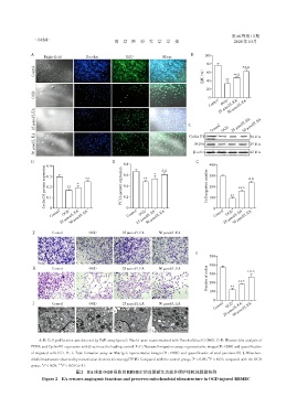
                 A,B:Cell proliferation was detected by EdU assay(green). Nuclei were counterstained with Hoechst(blue)(×200). C-E:Western blot analysis of
              PCNA and Cyclin D1 expression with β⁃actin as the loading control. F,G:Transwell migration assay:representative images(F,×200)and quantification
              of migrated cells(G). H,I:Tube formation assay on Matrigel:representative images(H,×100)and quantification of total junctions(I). J:Mitochon⁃
                                                                                        **
                                                                                 *
              drial ultrastructure observed by transmission electron microscopy(TEM). Compared with the control group,P < 0.05,P < 0.01;compared with the OGD
                         △△
                  △
              group,P < 0.05, P < 0.01(n=3).
                                 图2 EA恢复OGD损伤的RBMEC的血管新生功能并保护线粒体超微结构
                   Figure 2  EA restores angiogenic functions and preserves mitochondrial ultrastructure in OGD⁃injured RBMEC

