


Page 94 - 南京医科大学自然版
P. 94
第46卷第10期
               ·1524 ·                           南 京    医 科 大 学 学         报                        2026年10月


                      A                                            B
                           1.0                                          1.0
                          Fraction of positives  0.6                   Fraction of positives  0.6
                                                                        0.8
                           0.8

                           0.4
                                                                        0.4
                           0.2
                            0               Perfectly calibrated        0.2 0           Perfectly calibrated
                              00  0.2  0.4  0.6  0.8  1.0                 00   0.2  0.4  0.6  0.8  1.0
                                   Mean predicted probability                  Mean predicted probability
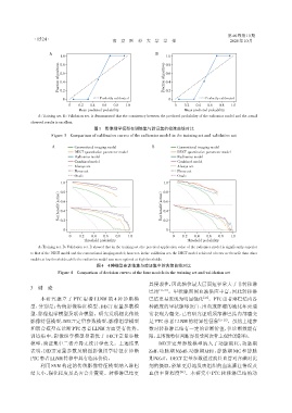
                 A:Training set. B:Validation set. It demonstrated that the consistency between the predicted probability of the radiomics model and the actual
              observed results is excellent.
                                         图3 影像组学模型在训练集与验证集的校准曲线对比
                       Figure 3 Comparison of calibration curves of the radiomics model in the training set and validation set


                       A         Conventional imaging model       B         Conventional imaging model
                                 DECT quantitative parameter model          DECT quantitative parameter model
                                 Radiomics model                            Radiomics model
                                 Combined model                             Combined model
                                 Always act                                 Always act
                                 Never act                                  Never act
                                 Orade                                      Orade
                           1.0                                         1.0
                          (action)  0.8                               (action)  0.8

                           0.6
                                                                       0.6
                          Net benefit  0.4                            Net benefit  0.4

                           0.2
                                                                       0.2
                            0                                           0
                              00   0.2  0.4  0.6  0.8  1.0                00   0.2  0.4  0.6  0.8  1.0
                                     Threshold probability                       Threshold probability
                 A:Training set. B:Validation set. It showed that in the training set,the practical application value of the radiomics model is significantly superior
              to that of the DECT model and the conventional imaging model;however,in the validation set,the DECT model achieved a better net benefit than other
              models at low thresholds,while the radiomics model was more optimal at high thresholds.
                                          图4 4种模型在训练集与验证集中的决策曲线对比
                          Figure 4 Comparison of decision curves of the four models in the training set and validation set

                                                                具侵袭性,因此轴位最大层面短径常大于非转移淋
              3  讨 论
                                                                巴结  [21-23] 。甲状腺周围血液供应丰富,因此转移淋
                  本研究建立了 PTC 患者 LLNM 的 4 种诊断模                   巴结更易表现为明显强化            [24] 。PTC患者淋巴结内各
              型,分别是:传统影像特征模型、DECT 定量参数模                         种浓度的甲状腺球蛋白、出血或肿瘤的液化坏死通
              型、影像组学模型及联合模型。研究发现相比传统                            常表现为囊变,已有研究证明颈部淋巴结内部囊变
              影像特征模型,DECT定量参数模型、影像组学模型                          是 PTC 患者 LLNM 的特异性征象         [21-23] 。虽然上述参
              和联合模型在诊断 PTC 患者 LLNM 方面更有优势。                      数对转移淋巴结有一定的诊断价值,但诊断效能有
              训练集中,影像组学模型显著优于 DECT 定量参数                         限,且图像特征判断容易受判读者主观性的影响。
              模型,验证集中二者差异无统计学意义。上述结果                                 DECT 定量参数模型纳入了动脉期 IC、动脉期
              表明,DECT 定量参数及碘图影像组学特征在诊断                          Zeff、动脉期 NZeff、动脉期λHU、静脉期 NIC 和静脉
              PTC患者LLNM转移中具有临床价值。                               期NZeff。DECT定量参数通过病灶血管对含碘对比
                  利用 SVM 构建的传统影像特征模型纳入淋巴                        剂的摄取,能够更好地反映组织的血流灌注情况及
              结大小、强化程度及是否合并囊变。转移淋巴结更                            血供丰富程度       [25] 。本研究中 PTC 转移淋巴结的动

