Page 14 - 南京医科大学自然版
P. 14

第44卷第5期
               ·602 ·                            南 京    医 科 大 学 学         报                        2024年5月


              A                                           B
                                           (%)  15  ***                IBMDM                   Control
                                                                                               S.T
                                                                 S.T
                                                                     -
                   S.T                     Cell containing ASC speck  10 5  GSK8612  -  + -  + +  Oligomer  1.5  S.T+GSK8612  ***
                                                                                                     ***
                                                                kDa
                                                                 100
                                                                                          1.0
                 ASC/DAPI merge             0  S.T+GSK8612     Insoluble+DSS  50  Dimer  Relative protein expression level  0.5 0  Oligomer Dimer Monomer
                                                                                               ***
                                                S.T
                   S.T+GSK8612                                 Soluble  25      Monomer
                                                                 25
                                                                                ASC
                                                                 35
                                                                                GAPDH


                 A:S.T(MOI:10)was used to infect IBMDM for 2 h,and endogenous ASC specks were detected by immunofluorescence(arrow). Scale bar:20 μm. B:
              After infecting IBMDM with S.T(MOI:10)for 2 h,insoluble samples crosslinked with DSS were taken for protein immunoblotting to detect oligomeriza⁃
                        **
                 *
              tion. P < 0.05,P < 0.01,and  *** P < 0.001(n=3).
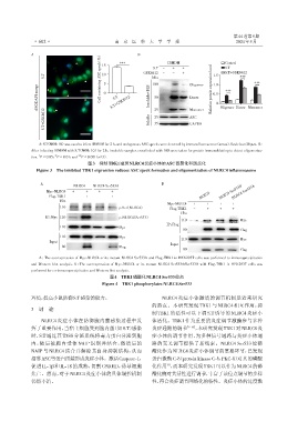
                                      图3 抑制TBK1减弱NLRC4炎症小体的ASC寡聚化和斑点化
                Figure 3 The inhibited TBK1 expression reduces ASC speck formation and oligomerization of NLRC4 inflammasome

                  A                                                 B
                                                                                                  NLRC4 Ser533A
                                NLRC4   NLRC4 Ser533A
                    Myc⁃NLRC4  +     +    +    +                                             NLRC4 Ser533D
                     Flag⁃TBK1  -    +    -    +                                    NLRC4
                          kDa
                                                                       Myc⁃NLRC4  +    +     +    +
                          110                      p⁃Ser(NLRC4)         Flag⁃TBK1  -   +     +    +
                                                                            kDa
                    IP:Myc 110                     p⁃NLRC4(Ser533)
                                                                            110                      Myc
                                                                       IP:Flag
                          110                      Myc
                                                                             80                      Flag
                          110                      Myc
                                                                            110                      Myc
                      Input
                                                                        Input
                           80                      Flag
                                                                             80                      Flag
                 A:The overexpression of Myc⁃NLRC4 or its mutant NLRC4 Ser533A and Flag⁃TBK1 in HEK293T cells was performed to immunoprecipitation
              and Western blot analysis. B:The overexpression of Myc⁃NLRC4 or its mutant NLRC4 Ser533A/Ser533D with Flag⁃TBK1 in HEK293T cells was
              performed for co⁃immunoprecipitation and Western blot analysis.
                                               图4 TBK1磷酸化NLRC4 Ser533位点
                                            Figure 4 TBK1 phosphorylates NLRC4 Ser533


              环境,提高小鼠防御S.T感染的能力。                                     NLRC4 炎症小体激活的调节机制是近来研究
                                                                的热点。本研究发现 TBK1 与 NLRC4 相互作用,抑
              3  讨 论
                                                                制 TBK1 的活性可以下调 S.T 诱导的 NLRC4 炎症小
                  NLRC4 炎症小体在防御胞内菌感染过程中发                        体活化。TBK1 作为重要的炎症调节激酶参与多种
              挥了重要作用,当宿主细胞受到胞内菌(如S.T)感染                         炎症通路的调节        [9-10] ,本研究发现TBK1对NLRC4炎
              时,S.T 通过其 T3SS 分泌系统将毒力蛋白分泌到胞                      症小体的调节作用,为多种信号通路与炎症小体通
              内,随后被胞内受体 NAIP 识别并结合,激活后的                         路的交叉调节提供了新线索。NLRC4 Ser533 位磷
              NAIP与 NLRC4 结合并解除其自身抑制结构,从而                       酸化作为NLRC4炎症小体调节的重要环节,已发现
              招募ASC等蛋白组装形成炎症小体。激活Caspase⁃1,                     蛋白激酶C⁃δ(protein kinase C⁃δ,PKC⁃δ)对其的磷酸
              促进 IL⁃1β和 IL⁃18 的成熟,切割 GSDMD,诱导细胞                 化作用 ,而本研究发现TBK1可以作为NLRC4的磷
                                                                      [3]
              焦亡。然而,对于NLRC4炎症小体的具体调控机制                          酸化酶对其活性进行调节,丰富了该位点调节的多样
              仍然不清。                                             性,符合炎症调节网络化的特性。炎症小体的过度激
   9   10   11   12   13   14   15   16   17   18   19