
Page 81 - 南京医科大学自然版
P. 81
第45卷第12期 侯超群,葛万里,彭云鹏,等. α⁃酮戊二酸通过XRCC3诱发急性胰腺炎发病的机制研究[J].
2025年12月 南京医科大学学报(自然科学版),2025,45(12):1756-1765 ·1763 ·
A B
NC vs. αKG heatmap_plot
2
1
0
P⁃value -1 Gene symbol <0.001
P
Myh9l1
-2
-lg Xrcc3 <0.001
Gpatch8 <0.001
Bzw1 <0.001
Nos3 <0.001
Spata31d1 <0.001
Pigw <0.001
log2 Fold Change
Rassf5 <0.001
Rpl26 <0.001
Ssna1 <0.001
NC1 NC2 NC3 αKG1 αKG2 αKG3
C D
1.5 NC αKG 1.5
** XRCC3 38 kDa **
Relative XRCC3 mRNA expression 1.0 β⁃actin 42 kDa Relative XRCC3 protein expression 1.0
0.5
0.5
0.0 0.0
NC αKG NC αKG
E XRCC3 (%) 50 F OE⁃XRCC3 0.4 * G 0.5 *
(×400) 40 ** XRCC3 NC 38 kDa 0.3 0.4
0.3
NC Relative ratio of XRCC3 prositive stained area 30 β⁃actin 42 kDa Relative XRCC3 protein expression 0.2 Cell viability rate 0.2
20
0.1
0.1
(×400) 10 0 0.0 NC 0.0 αKG
αKG NC αKG OE⁃XRCC3 αKG+OE⁃XRCC3
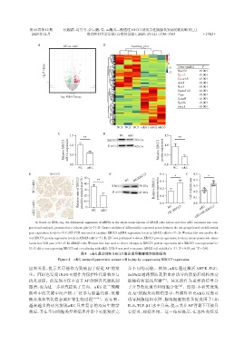
A:Based on RNA⁃seq,the differential expression of mRNA in the whole transcriptome of AR42J cells before and after αKG treatment was com⁃
pared and analyzed,presented as a volcano plot(n=3);B:Cluster analysis of differentially expressed genes between the two groups based on differential
gene expression levels(n=3).C:RT⁃PCR was used to examine XRCC3 mRNA expression levels in AR42J cells(n=3);D:Western blot was used to de⁃
tect XRCC3 protein expression levels in AR42J cells(n=3);E:IHC was performed to detect XRCC3 protein expression levels in mouse pancreatic tissue
(scale bar=100 μm,n=6);F:In AR42J cells,Western blot was used to detect changes in XRCC3 protein expression after XRCC3 overexpression(n=
**
*
3);G:After overexpressing XRCC3 and co⁃culturing with αKG,CCK⁃8 was used to measure AR42J cell viability(n=3). P < 0.05 and P < 0.01.
图4 αKG通过抑制XRCC3表达诱导胰腺腺泡细胞损伤
Figure 4 αKG induced pancreatic acinar cell injury by suppressing XRCC3 expression
因果关系,提示其可能作为致病因子促进 AP 的发 全不同的功能。例如,αKG 通过激活 AMPK⁃PGC⁃
生。同时也发现INDO可能作为保护性代谢物参与 1α/Nrf2 通路预防高脂血症诱导的脂肪肝线粒体功
抗炎过程。此发现不仅丰富了AP 的潜在代谢机制 能障碍和氧化应激 [25] 。其还能作为重要的信号分
图谱,也为进一步研究提供了方向。αKG是三羧酸 子介导免疫调节和细胞分化 [26] 。然而,本研究发现
循环中的关键中间产物,广泛参与能量代谢、氨基 在 AP 细胞及动物模型中,外源性补充αKG 反而可
酸合成和氧化稳态调控等生物过程 [23-24] 。近年来, 诱导胰腺组织水肿、腺泡细胞损伤及促炎因子(如
越来越多的研究发现αKG 具有复杂的双向生物学 IL⁃6、TGF⁃β1)水平升高,提示其在AP背景下可能具
效应,其在不同细胞类型和病理背景中可能发挥完 有促炎、致病作用。这一结果提示,在急性炎症反