


Page 31 - 南京医科大学自然版
P. 31
第45卷第7期               沈埝萱,孙陈晨,孔灵杰,等. ST3GAL3在食管鳞癌中的作用与机制研究[J].
                  2025年7月                    南京医科大学学报(自然科学版),2025,45(7):925-935,944                     ·929 ·


                  A          GSE130078(n=23)       B          GSE111011(n=7)       C         GSE119436(n=4)
                                  ***                              *                              **
                       5 4                             5 4                            5
                      (normalized counts)  3 2        (normalized counts)  3 2       (normalized counts)  4


                      log2  1                         log2  1                        log2  2

                       0                               0                              0
                            Normal     Tumor                Normal     Tumor               Normal     Tumor
                   A-C:The mRNA expression levels of ST3GAL3 in paired ESCC and adjacent normal tissues based on the expression profile of GSE130078(A,
                                                          **
                                                   *
                n=23),GSE11011(B,n=7),and GSE119436(C,n=4). P < 0.05,P < 0.01 and  *** P < 0.001.
                                          图1 ST3GAL3 mRNA水平在ESCC临床标本中高表达
                         Figure 1 The expression of ST3GAL3 mRNA was highly expressed in ESCC clinical sample tissues

                A        4        ***              B            Normal tissues               Tumor tissues

                      Relative mRNA expression  (fold change) 3 2 1  Patient #1  × 100  × 200  × 100   × 200


                         0
                             Normal   Tumor
                C
                    1.0                              Patient #2
                   Accumulated survival rate  0.6  ST3GAL3 (n=66) low                                      100 μm
                    0.8

                                     ST3GAL3 (n=31)
                    0.4
                                     high
                    0.2
                     0  log rank P=0.002
                        0  10  20  30  40  50  60
                             Survival time(months)
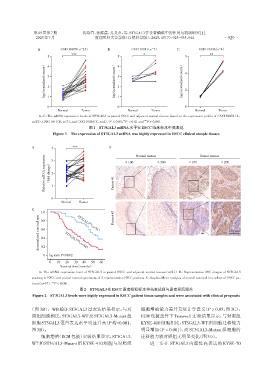
                   A:The mRNA expression level of ST3GAL3 in paired ESCC and adjacent normal tissues(n=21). B:Representative IHC images of ST3GAL3
                staining in ESCC and paired normal specimens of 2 representative ESCC patients. C:Kaplan⁃Meier analysis of overall survival in a cohort of ESCC pa⁃
                tients(n=97).  *** P < 0.001.
                                      图2 ST3GAL3在ESCC患者组织标本中高表达且与患者预后相关
                Figure 2  ST3GAL3 levels were highly expressed in ESCC patient tissue samples and were associated with clinical prognosis


               (图 3B)。WB验证ST3GAL3过表达结果显示,与对                       细胞增殖能力差异无统计学意义(P > 0.05,图3C);
                照组细胞相比,ST3GAL3⁃WT及ST3GAL3⁃Mutant组                 ECM包被条件下Transwell实验结果显示,与对照组
                细胞ST3GAL3蛋白表达水平明显升高(P均<0.001,                     KYSE⁃410细胞相比,ST3GAL3⁃WT组细胞迁移能力
                图3B)。                                             明显增加(P < 0.001),而ST3GAL3⁃Mutant组细胞的

                    细胞增殖(ECM包被)实验结果显示,ST3GAL3⁃                    迁移能力较对照组无明显变化(图3D)。
                WT 和 ST3GAL3⁃Mutant 组 KYSE⁃410 细胞与对照组                 进一步在 ST3GAL3 内源性高表达的 KYSE⁃70

