
Page 36 - 《南京医科大学学报》自然科学版2026年第2期
P. 36
第46卷第2期
·192 · 南 京 医 科 大 学 学 报 2026年2月
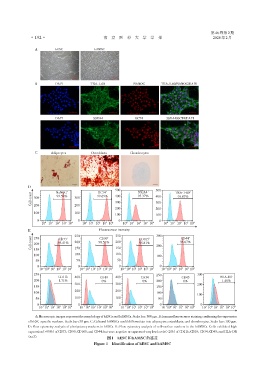
A hESC hAMSC
B DAPI TRA⁃1⁃60 NANOG TRA⁃1⁃60/NANOG/DAPI
DAPI SSEA4 OCT4 SSEA4/OCT4/DAPI
C Adipocytes Osteoblasts Chondrocytes
D
500 SSEA4 + 500 TRA⁃1⁃60 +
+
+
NaNOG
OCT4
Cell count 300 99.59% 300 99.69% 400 95.37% 400 99.07%
300
300
200
200
200 200
100 100
100 100
0 0 0 0
0
3
2
3
0
0
3
2
2
1
3
2
0
10 10 1 10 10 10 4 10 10 10 10 10 4 10 10 1 10 10 10 4 10 10 1 10 10 10 4
E 250 Fluorescence intensity 300
250
Cell count 250 95.41% + 200 98.51% + 200 95.81% + 200 96.67% +
CD44
CD90
CD73
CD105
200
150
150
150
100 100 100 100
50 50 50
0 0 0 0
2
2
0
0
-1
1
1
3
-1
3
0
-1
2
2
3
-1
1
1
0
3
10 10 10 10 10 10 4 10 10 10 10 10 10 4 10 10 10 10 10 10 4 10 10 10 10 10 10 4
250 250 300
CD11b 400 CD19 400 CD34 CD45 HLA⁃DR
200 1.71% 0% 0% 200 0% 1.48%
300 300 200
150 150
200 200
100 100
100
50 100 100 50
0 0 0 0 0
2
2
3
3
0
1
1
-1
-1
0
3
3
-1
0
2
-1
0
1
1
0
-1
2
1
2
3
10 10 10 10 10 10 4 10 10 10 10 10 10 4 10 10 10 10 10 10 4 10 10 10 10 10 10 4 10 10 10 10 10 10 4
A:Microscopic images represent the morphology of hESCs and hAMSCs. Scale bar:500 μm. B:Immunofluorescence staining confirming the expression
of hESC⁃specific markers. Scale bar:50 μm. C:Cultured hAMSCs could differentiate into adipocytes,osteoblasts,and chondrocytes. Scale bar:100 μm.
D:Flow cytometry analysis of pluripotency markers in hESCs. E:Flow cytometry analysis of cell⁃surface markers in the hAMSCs. Cells exhibited high
expression(>95%)of CD73,CD90,CD105,and CD44,but were negative or expressed very low levels(<2%)of CD11b,CD19,CD34,CD45,and HLA⁃DR
图1 hESC和hAMSC的鉴定
(n=3).
Figure 1 Identification of hESC and hAMSC