


Page 24 - 南京医科大学自然版
P. 24
第46卷第7期
               ·970 ·                            南 京    医 科 大 学 学         报                        2026年7月


              物 MAP⁃2 进行染色,发现 DAPI 所标记的所有神经
              元细胞核都被MAP⁃2所包绕,说明分离出来的细胞
              就是原代神经元,此外,该系列细胞形态良好,核大
              而清晰(图 6),为后续神经元相关实验提供了前期
              基础。
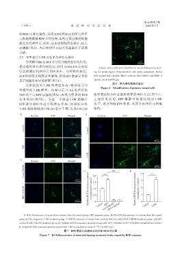
              2.5  BPF通过3⁃HK引起原代神经元损伤
                  为明确3⁃HK在BPF介导的突触损伤中的作用,
                                                                                              50 μm
              通过体外培育原代神经元,利用 DCFH⁃DA 荧光探
                                                                    Primary neural cells were identified by immunofluorescence stain⁃
              针法检测原代神经元 ROS 水平。与对照组相比,                          ing for morphological characterization and purity assessment. Nuclei
              BPF 组的荧光强度显著增强,表明 BPF 暴露显著促                       were stained with Hoechst(blue);neurons were labeled with MAP ⁃ 2
              进了细胞内ROS的积累(图7A)。                                 (green). Scale bar=50 μm.
                                                                             图6 原代神经细胞的鉴定
                  为单独探究 3⁃HK 的毒性作用,将神经元与
                                                                     Figure 6  Identification of primary neural cells
              外源性的 3⁃HK 孵育。结果显示,3⁃HK 处理组的
              ROS 水平与 BPF 暴露组相似,表现为明显的 ROS                      能显著逆转 BPF 暴露所诱导的 ROS 升高(图 7C)。
              水平增加(图 7B)。为进一步验证 3⁃HK 通路在                        上 述 结 果 表 明 ,BPF 暴 露 可 能 通 过 增 加 3⁃HK
              BPF 诱导 ROS 生成中的潜在作用,使用特异性                         水 平,诱导细胞 ROS 积累,从而导致神经元细胞

              3⁃HK 通路抑制剂 GSK180 进行干预,发现 GSK180                  损伤。
               A                                           B
                      Hochest      ROS         Merged              Hochest          ROS           Merged

                 Control                                     Control


                                                   50 μm
                                                                                                         50 μm
                 BPF

                                                             3⁃HK
                                                   50 μm
               C
                      Hochest      ROS         Merged
                 Con+GSK180                              D      2.0    ***  ***          E      4     ***  50 μm


                                                   50 μm        1.5                             3
                 BPF+GSK180                                  Relative ROS fluorescence intensity (normalized to Control)  1.0  Relative ROS fluorescence intensity (normalized to Control)  2 1


                                                                0.5

                                                   50 μm         0 Control Con+GSK180           0 Control  3⁃HK
                                                                        BPF
                                                                          BPF+GSK180

                 A:ROS fluorescence of mouse brain neurons from the control group,BPF exposure group. B:The ROS fluorescence,in neurons from the control
              group and the exogenous 3⁃HK treatment group. C:ROS fluorescence of mouse brain neurons from the control with GSK180 treatment group,and BPF
              combined with GSK180 treatment group. D:Relative ROS fluorescence intensity treated with BPF,GSK180,or BPF+GSK180(normalized to Control).
              E:Relative ROS fluorescence intensity treated with 3⁃HK(normalized to Control).  *** P < 0.001(n=3).
                                            图7 BPF暴露引起神经元损伤的ROS荧光图
                            Figure 7  ROS fluorescence of neuronal damage in mouse brain caused by BPF exposure

