


Page 50 - 南京医科大学自然版
P. 50
第46卷第9期
               ·1320 ·                           南 京    医 科 大 学 学         报                        2026年9月


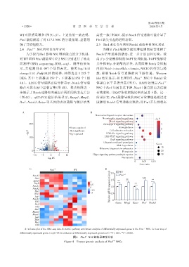
              WT 对照明显增多(图3C、D)。上述结果一致表明,                        高度一致(图 4D),提示 Notch 信号通路可能介导了
              Pin1 敲除解除了对 C17.2 NSC 的分裂限制,显著增                   Pin1缺失引起的增殖表型。
              强了其增殖能力。                                          2.5  Pin1通过负向调控Notch1通路抑制NSC增殖
              2.4  Pin1 -/-  NSC的转录组学分析                              为确证 Pin1 敲除引起的增殖增强是否依赖于
                  为了探究Pin1影响NSC增殖能力的分子机制,                       Notch 信号通路的激活,进一步开展回补实验。使
              对 WT 型和 Pin1 敲除型 C17.2 NSC 分别进行了转录                用了γ⁃分泌酶抑制剂DAPT处理细胞,DAPT能够特
              组测序(RNA sequencing,RNA⁃seq)。测序结果显                 异性抑制γ⁃分泌酶的活性,从而阻断 Notch 受体胞

              示,共检测到 41 097 个基因表达。按照|log2 (fold                 内段(Notch intracellular domain,NICD)的剪切与释
              change)|≥1,Padj≤0.05 的标准,共筛选出 1 215 个             放,抑制 Notch 信号通路的向下游传递。Western
              DEG,其中上调基因 359 个,下调基因 856 个(图                     blot 结果显示,相比 WT 组,Pin1 NSC 中 Notch1 受
                                                                                             -/-
              4A)。KEGG 信号通路富集分析显示,Notch 信号通                     体蛋白水平显著升高(图 5)。DAPT 处理后 Pin1                 -/-
              路在差异基因中显著富集(图 4B)。聚类热图进一                          NSC 中 Hes1 基因表达下降,Notch1 蛋白的高表达被
              步展示了Notch通路相关基因在两组间的表达差异                          有效逆转,且Ki67阳性细胞比例也显著下降。这一
             (图 4C)。qRT⁃PCR 验证结果显示,Ramp1、Mmp2、                  结果证实,Pin1敲除导致的NSC异常增殖是通过过
              Hes1、Notch3、Rictor 等基因的表达趋势与测序结果                  度激活Notch1信号通路实现的,即Pin1在生理状态


                A                                            B
                                                                Neuroactive ligand⁃receptor interaction
                                                                    Neurotrophin signaling pathway     -lg P
                                                                         Notch signaling pathway
                    200                                              NF⁃kappa B signaling pathway          3
                                               Down⁃regulated                  Axon guidance
                                                                                                           2
                    P                          Not⁃significant           Cell adhesion molecules
                    -lg                        Up⁃regulated            PI3K⁃Akt signaling pathway          1
                                                                      JAK⁃STAT signaling pathway
                    100
                                                                         Rap1 signaling pathway
                                                                     Ubiquitin mediated proteolysis
                                                                                                        Count
                                                                             RNA degradation
                                                                                                           10
                                                                  Ribosome biogenesis in eukaryotes
                      0                                                          Ferroptosis               20
                                                              Hippo signaling pathway⁃multiple species     30
                       -10  -5    0    5    10
                                log2 FC                                          Necroptosis
                                                                                         0    0.05  0.10
                                                                                             Gene ratio
                    C             Pin1 -/-       WT                   D         WT
                       Notch3                                             2.0   Pin1 -/-
                        Hes1                                                                   ***
                       Aph1b                                3             1.5  **   **               **
                        Dtx3l                                                            **
                       Notch1                               1.5          Relative mRNA expression levels  1.0
                       Notch2                               0
                        Hes5                                -1.5
                        Hey1                                -3            0.5
                        Jag1
                       Notch4                                              0
                        Dll1                                                 Alcam  Mmp2 Rictor  Hes1 Notch3
                        Hes2
                        Dll3
                        Hes7
                       Maml3
                       Maml1
                        Jag2
                        Hes6
                        Hes3
                        Dll4
                                                                                               -/-
                 A:Volcano plot of the RNA⁃seq data. B:KEGG pathway enrichment analysis of differentially expressed genes in the Pin1 NSC. C:Heat map of
                                                                          **
                                                                                 ***
              differentially expressed genes. D:qRT⁃PCR validation of differentially expressed genes(n=3). P < 0.01, P < 0.001.
                                                 图4 Pin1 NSC的转录组学分析
                                                         -/-
                                                                           -/-
                                           Figure 4 Transcriptomic analysis of Pin1 NSCs

